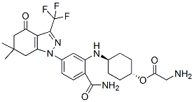
This problem also calls for the development of highly efficient laboratory models of HPV infection. Altogether, five of the putative papillomavirus microRNAs were encoded by HPV 16, one by HPV 38, one by HPV 68, one by HPV 45 and one by HPV 6. HPV 68 belong to alpha-papillomaviruses, whereas HPV 38 is a beta-papillomavirus. None of the candidate HPV encoded microRNAs had similarity to known human microRNA sequences. Of the validated microRNAs, HPV16-miR-H1-1 is located within the E1 region of the coding strand, and HPV16-miR-H2-1 in the negative strand corresponding to the LCR region. Intriguingly, the HPV16-miRH2-1 sequence is present in a number of HPV 16 isolates but has a one-nucleotide deletion in the prototype sequence. Many of the isolates have been cloned from carcinoma tissues, suggesting that the ability to express this particular microRNA might promote carcinogenesis. Interestingly, the Orbifloxacin pre-microRNA sequence of HPV38-miR-H1, encoded by the E7 region, is shared by HPV types 22, 23, 120, 104 and 115, which are all members of the betapapillomavirus genus. Moreover, the pre-miRNA sequence of HPV45-miR-H1, encoded by the L1 region, is partially similar to HPV 16, suggesting Mepiroxol evolutionary divergence of viral miRNA function between HPV types. Although the deep sequencing read counts for the HPV 6 encoded miRNA were high, we were not able to validate it by qPCR, possibly due to the specific design of TaqMan assays. Because of the very short length of the miRNA there are very limited possibilities to alter the assay design if no results are obtained with the qPCR test. Several of the putative miRNA sequences were encoded by the negative DNA strand, which disagrees with the consensus that all papillomavirus transcripts originate from the positive strand of papillomavirus genomes. Although in this first report we did not study the mechanisms of transcription of HPV microRNAs, our methodology should practically exclude RNA degradation products or DNA from sequencing libraries. Viral miRNAs may also possess features that do not follow the canonical properties of human miRNAs. The precursor sequence of HPV16-miR-H1 is still uncertain and needs further validation of length and exact sequence. Due to low expression level of this miRNA we were not able to establish its exact length. Merkel cell polyomavirus encoded MCV-miR-M1-5p, which was first predicted from VMir and validated, and further identified by Illumina sequencing and validated by qRT-PCR, has a 59end 2-nt shift from the VMir predicted MCV-mir-M1 mature sequence, which has also been shown to exist and  be functional in vitro. Further studies are needed to prove whether the isomiRs presented here could also exist and be functional under some conditions. Entire tissue samples consisting of both healthy and infected cells were used for these studies. Robust signals were seen in cervical tissue in in situ hybridization, often colocalizing with and restricted to regions staining for p16INK4a, which is considered a surrogate marker for high-risk HPV oncogene activity. In situ hybridization also showed altered distribution of human miR-205, whose high expression has been reported before in CaSki cells and in cervical cancer tissues. miR-205 was also recently shown to promote proliferation of human cervical cancer cells. Although some viral miRNAs are occasionally expressed at high levels, low level of expression has also been shown biologically relevant, for example for Merkel cell polyomavirus miRNA. Those authors speculated that even low levels of viral miRNA expression might be sufficient to regulate host immune response. However, the signals in the in situ assays for the U6, miR-205 and HPV miRNAs cannot be directly compared as a measure of the expression level.