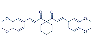
Endothelial dysfunction might be due to uncoupled eNOS. Several studies have shown that relaxin effects are endothelium and NO dependent. Thus, impaired endothelium mediated vascular relaxation in the dTGR model could explain the missing relaxin effectiveness. Recently Parikh et al. showed beneficial effects of relaxin on Eleutheroside-E atrial fibrillation in spontaneously hypertensive rats. Relaxin treatment reversed the transcripts for fibrosis, increasing conduction velocity, reduced electrophysiological abnormalities, and reversed atrial hypertrophy. In their study a one-week therapy was ineffective in suppressing atrial  fibrillation and longer relaxin treatment was necessary. The authors speculate that reversal of fibrosis is a slow process as a result of the slow collagen turnover rate of around 5% per day in healthy hearts. The fibrosis in our dTGR is much more pronounced. This state-of-affairs and the fact that fibrosis accelerates over time might be the reason why relaxin was not successful in ameliorating target-organ damage in dTGR. We were also interested in the kidney in dTGR. Recently Yoshida et al demonstrated that relaxin protected against ischemia/reperfusion-induced renal injury by reducing apoptosis and inflammation. Relaxin also preserved renal function in their model. Their relaxin dose, namely 500 ng/h was higher than our high-dose. Moreover, the design of their study was based on acute renal failure; our study was chronic in nature. Parikh et al did not investigate inflammation, while Yoshida et al showed that the TNFa receptor-1 was up regulated in the kidney and normalized by relaxin. We found no such effects in dTGR model. The transcription factors nuclear factor-kB and activator-protein 1 are strongly activated and responsible for the sustained inflammatory and proliferative response in this model. Macrophages, dendritic cells and CD4 and CD8 T cells are strongly activated. Surface adhesion molecule expression, such as ICAM-1, VCAM-1, TNFa and interleukin-6, tissue factor production, and activation of enzymes producing reactive oxygen species are highly induced. We speculate that relaxins anti-inflammatory potential was not sufficient to counteract the pro-inflammatory storm in dTGR. In contrast to our findings, Lekgabe et al. showed that relaxin reduced target-organ damage in spontaneous hypertensive rats. However, end-organ damage takes 9�C10 months to develop in that model and is far less severe than the effects reported here. Relaxin, applied for 2 weeks, normalized fibrosis in heart and kidney, inhibited cell and increased MMP-2 expression. Blood pressure was not affected by relaxin treatment and mortality was not investigated. Wong et al investigated the effects of relaxin on fibrosis in streptozotocin -treated transgenic mRen-2 rats. That model is also Ang II-mediated and features fairly severe changes. Relaxin did not ameliorate glomerulopathy in this accelerated model of type 1 diabetes. Relaxin did not reduce Saikosaponin-B2 hypertension or albuminuria. Similar to our study, the authors were puzzled by the negative results. The authors observed that relaxin was very successful in reversing fibrosis when the TGF-b1 pathway was activated and subsequently phosphorylation and down regulation of smad-2 occurred. This was not the case in the dTGR.