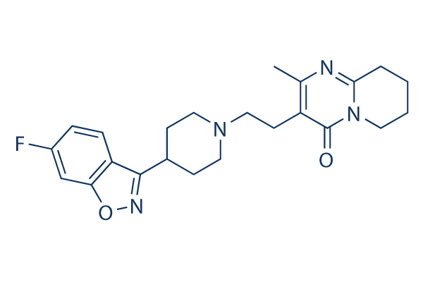
For example, the reduction in IGF-IR activation by the binding of specific antibodies leads to apoptosis of cancer cells. A recent study using the monoclonal antibody ‘Figitumumab’, supported the potential therapeutic efficacy of anti-IGF-IR strategies for the treatment of patients with Ewing’s sarcoma. However several drug companies have recently stopped development of drugs designed to block IGF-R signaling, expressing frustration over the ineffectiveness of drugs that have been developed, blaming the biological complexity of the IGF system. Based on the hard won findings, it is now clearly apparent that a ‘systems approach’is needed to understand why a single drug target may be ineffective for managing IGF-IR signaling. Indeed, it points to the fact that several drugs acting together may be required to effectively block a signaling pathway. From a sensitivity analysis for our model, we find that it is likely that negative feedback processes act to neutralize the effect of attempting to block a single target. It is expected that treatment of patients with a variety of disease processes in all tissues of the body may be enhanced when there is an improved understanding of the processes that regulate the cell’s exposure to IGF within a tissue from a circulating source of IGF. To contribute towards this goal, this paper is focused developing a systems model to estimate the free and total IGF concentrations within a single tissue �C articular cartilage. Cartilage was chosen primarily because we are aware of appropriate data to enable calibration of the model. IGF-I and IGF-II bind to the IGF-IR receptor, leading to activation of a receptor tyrosine Sipeimine kinase and subsequent downstream signaling via the AKT pathway. The strength of activation of the signaling depends on the fraction of overall receptors that have formed a complex with their ligands. However the downstream pathway activation need not be proportional to the receptor occupancy. For example, previous studies on cartilage have shown there is  a certain threshold of IGF-I/ IGF-IR concentration that needs to be exceeded before protein synthesis is activated. In this paper IGF-IR complex formation is included, but no downstream signaling processes are modeled, so the IGF-IR complex concentration is adopted here as the primary biological marker of functional activity due to IGFs in the tissue. Although IGF-II is widely argued to play an important role in embryonic and foetal life, recent studies indicate that IGF-II is also important in adults for muscle, brain and other tissues by signaling through the receptor IGF-IR. While many tissues produce IGF-II, most tissues produce little or no IGF-I, with the majority of IGF-I in tissues originating from production by the liver. Only IGF-II binds to the IGF-IIR receptor. Formation of IGF-IIR complexes usually has no known downstream signaling consequences, although it has been reported that binding of IGF-II to IGF-IIR may provide a possible mechanism for the regulation of cardiomyocyte apoptosis. 4-(Benzyloxy)phenol Instead it is thought that the primary role of the IGF-II-IGF-IIR complex is the regulation of the IGF-II concentration in the tissue, i.e via sequestration and removal of the IGF-II-IGF-IIR complex through lysosomal degradation. In other words, IGF-IIR is postulated to be a ‘clearance receptor’. One conceivable mechanism for regulating the IGF-IR receptor complex concentration in the tissue is to regulate the ratio of IGF-I and �CII in the tissue, as IGF-I and �CII ligands competitively bind to IGF-IR. Note the IGF-II concentration in human plasma is typically three-fold higher than that of IGF-I, and the ratio of IGF-II/IGF-I may reach over 300 in a tumor.