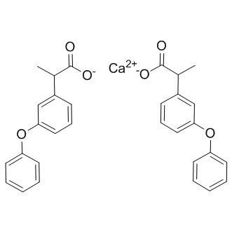
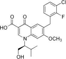
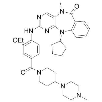
HIFs are heterodimeric transcription factors which have two structurally related subunits, an Orbifloxacin oxygen sensitive HIFa subunit and a constitutively expressed HIF? or ARNT subunit. Sox2 is important for pulmonary branching morphogenesis, epithelial cell differentiation and is exclusively expressed in the proximal parts of the lung. However, in mycHIF3a expressing lungs, Sox2 is present in epithelial cells of both proximal airways and certain alveoli at postnatal day 1, suggesting that Hif3a is able to induce proximal cell fate. The basal cell marker p63 is expressed in the esophagal and tracheal epithelium, and previously we showed that ectopic Sox2 expression induced the appearance of p63 positive cells in the epithelium of the bronchioles and Folinic acid calcium salt pentahydrate enlarged distal airspaces. Therefore, we analysed  the distribution of basal cells in the mycHIF3a expressing lungs and found that p63 is abnormally expressed in the alveolar epithelial cells of mycHIF3a expressing lungs, contrasting the unique expression in the trachea. Hypoxia inducible factors are an important family of proteins involved in the regulation of the cellular response to hypoxia. Its functions are required from the earliest steps of mammalian life to the correct development of multiple organs and tissues, like the placenta, trophoblast formation, bone development, heart and vascular development. The importance of the hypoxia response was shown by the identification of human mutations in the VHL-HIF pathway in different diseases. Gene ablation studies in mice have revealed in more detail the specific and important roles of the different subunits of the Hifa/Hif? heterodimers. Inactivation of the stable subunit, Hif1?, resulted in severe embryonic defects and premature death. The disruption of the different Hifa genes identified specific roles for the individual Hifa isoforms. Hif1a knockout mice die early at gestation, have multiple developmental defects in neural tubeforrmation, vascularization, heart development, neural crest migration, whereas depending on the genetic background of the mouse strain, Hif2a knockout out mice ranging from early embryonic lethality to adulthood. Early-life stressors such as maternal undernutrition, overnutrition, hypercholesterolemia, corticosteroid therapy, uteroplacental insufficiency, or hypoxia program metabolic adaptations that initially favor survival but are ultimately detrimental to adult health. In laboratory rodents, low-protein diet during gestation and lactation has been known to reduce the life expectancy of offspring. The maternal protein restriction in the rat model of In Utero Protein Restriction is one of the most extensively explored model. The low-protein fed mothers give birth to growth-restricted offspring,, and when suckled by their mothers maintained on the same low-protein diet, they remain permanently growth-restricted, despite being weaned on a normal diet. Also, early-life undernutrition is associated with higher blood tryptophan levels, brain serotonin and impairment of the serotonergic control of feeding in female adult rats. Recently, we have shown that circadian clock of the hypothalamus is altered in young rats subsequently to perinatal undernutrition, however there is no proof that this dysregulation exists in other tissues as well. In rodents, the emergence of circadian clock outputs occur during the first 2 or 3 weeks after birth.
Month: June 2019
In contrast with the results that both TLR1 and TLR4 were up-regulated with normal after combination chemotherapy
Furthermore, we observed an increased nuclear S100A15 3,4,5-Trimethoxyphenylacetic acid expression in lung cancer tissues not only in stage IV NSCLC compared to stage IIIB NSCLC, but also in the patients with stable or progressive disease in comparison to those with a partial response after first line combination chemotherapy with CDDP and GEM. Additionally, a high percentage of S100A15 nuclear Tulathromycin B stained cells was the only independent factor associated with three-year overall survival. This suggests that nuclear accumulation of S100A15 may be linked to metastasis potential, treatment response, and long-term outcomes. In accordance with the results published in the Human Protein Atlas website,  we found up-regulation of S100A15 in NSCLC cancer tissues compared to normal lung tissues surrounding the tumor. Nonetheless, S100A15 in either cancer tissues or PBMC could not distinguish AC from SCC in this small sample-size study. Additionally, quantitative RT-PCR result for S100A15 was positively correlated with cytoplasmic S100A15 staining intensity score but not with other parameters in IHC staining assessment, implying that cytoplasmic protein expression rather than nuclear protein expression may affect S100A15 expression in peripheral immune cells, such as monocyte. Disruption of the calcium signaling pathway, such as by the S100 family, has been implicated as a central mechanism in tumorigenesis, specifically tumor invasion and metastasis. Both S100A15 and S100A7 proteins have been demonstrated to be distinctly expressed in normal breast tissue and breast cancer. Since S100A15 was found to be chemotactic for both granulocytes and monocytes, and to act synergistically with highly homologous S100A7 to enhance inflammation, both proteins could influence lung tumor progression. Additionally, E. coli can modulate the human S100A15 expression of keratinocytes by recognition through TLR4, suggesting that S100A15 may play a role in innate immunity. Selective expression of S100A7 in lung SCC and large cell carcinomas has been demonstrated, but not in AC or small cell carcinomas. Nuclear accumulation of S100A7 has been reported to be associated with a poor prognosis in head and neck cancer. To the best of our knowledge, the current study is the first to demonstrate that nuclear accumulation of S100A15 may be linked to an increased risk of distant metastasis, and that S100A15 may serve as a candidate biomarker for predicting treatment response and survival. However, large scale longitudinal studies are warranted to evaluate the potential of S100A15 as a determinant of advanced tumor stage and/or a predictor of long-term outcomes in NSCLC. Stimulation with TLR7 agonists on human lung cancer cells has been shown to lead to increased tumor cell survival and chemoresistance. On the other hand, systemic administration of TLR7 agonists has also been found to induce significant antitumor activity, which could be potentiated by cyclophosphamide. In a cell culture model, TLR7 agonists were found to enhance tumor cell lysis by human gamma delta T cells. Taken together, these findings suggest that enhancing the TLR7 expression in immune cells may potentiate the antitumor effect of combination chemotherapy in advanced stage NSCLC patients.
Putative redox enzyme that converts the hydroxymycolate products of MmaA4 into keto-mycolic acids
While these models are attractive, confirmation will require further study and more information on the structure of the proteins, their mode of interaction and how the drugs alter these interactions. Peroxisomes are single-Lomitapide Mesylate membrane bound, multifunctional and highly Tulathromycin B dynamic organelles of most eukaryotic cells, which fulfil important metabolic functions in hydrogen peroxide and lipid metabolism. Their function has also been linked to developmental processes, stress response, age-related disorders, and antiviral innate immunity. Remarkably, the peroxisomal compartment shows high plasticity and responds to developmental, environmental, and metabolic stimuli with alterations in organelle number, morphology and protein content. Peroxisomes can multiply by growth and division of pre-existing organelles or, as particularly demonstrated in yeast, can form de novo from the endoplasmic reticulum. Whereas considerable progress has been made in the identification of key factors involved in these processes, the underlying mechanisms and the regulation of these processes are only poorly understood. The assembly of peroxisomes and protein import into the organelle requires the action of essential proteins, so called peroxins, which are encoded by PEX genes. Mutations in many PEX genes have been identified as the cause of severe and often lethal peroxisome biogenesis disorders. Peroxisome formation by growth and division involves the deformation and elongation of the peroxisomal membrane, its constriction and final scission. Similar  to de novo biogenesis from the ER, growth and division of peroxisomes follows a multistep maturation pathway, which results in the formation of new daughter peroxisomes. In mammals, Pex11 proteins are so far the only proteins discovered capable of deforming and elongating the peroxisomal membrane. Hence, the mechanistic details of peroxisomal growth and division and the individual functions of the human Pex11 proteins have attracted great attention as they have been linked to new disorders affecting peroxisome morphology and dynamics. It has recently been reported that Pex11 proteins feature amphipathic helices that can insert into the peroxisomal membrane, thus influencing membrane bending. In line with this, Pex11 proteins are suggested to reorganize the peroxisomal membrane prior to fission,, and to mediate interactions with the peroxisomal fission machinery. The machinery for membrane scission includes the membrane adaptor proteins Fis1 and Mff, which are involved in the recruitment of the dynamin-like large GTPase DLP1/Drp1 to constriction sites on the peroxisomal membrane. DLP1 is supposed to assemble in spiral-like structures around constricted membranes to mediate membrane scission through GTP hydrolysis leading to the formation of new peroxisomes. Interestingly, mitochondria and peroxisomes, which are metabolically linked to each other, share these key components of their division machinery supporting a closer interorganellar relationship,,, whereas Pex11 proteins are exclusively peroxisomal. Pex11 proteins are conserved amongst species; however, many organisms contain various ����isoforms���� which are poorly characterized on a functional level, and may differ in their biochemical properties. Furthermore, their membrane topology is not entirely clear and may vary amongst different species. The mammalian genome encodes for three Pex11 proteins, Pex11pa, Pex11pb, and Pex11pc, which are thought to be integral membrane proteins with their N- and C-termini facing the cytosol.
Macrophage colony-stimulating factor and interleukin followed by induction of DC maturation
Using a proinflammatory cytokine cocktail composed of tumor necrosis factor -a, IL-1b, IL-6 and prostaglandin E2. Over the years, it has become apparent that these “gold-standard” DCs, commonly referred to as ‘IL-4 DCs’, are suboptimal in terms of antigen presentation function and T cell stimulatory capacity. This explains the impetus behind the many efforts that are currently being made to optimize the culture conditions for ex vivo monocyte-derived DC generation. Within this context, we and others have shown that the immunostimulatory properties of monocyte-derived DCs can be significantly enhanced by replacing IL-4 with IL-15 for DC differentiation and by using Toll-like receptor stimuli to trigger DC maturation. In addition, we have found that these so-called ‘IL-15 DCs’ display a rather unconventional DC phenotype, with a subset of these cells being positive for the cell surface marker CD56. Since CD56 is the archetypal phenotypic marker of NK cells, we here aimed to investigate whether IL-15 DCs also bear functional resemblance with NK cells in terms of cytotoxic activity. In this study, IL-15 DCs are shown to possess potent tumor antigen presentation function in combination with lytic potential against the classical NK cell target cell line K562, thus confirming the hypothesis that IL-15 DCs qualify for the designation of killer DCs. To further address the possibility that the observed lytic activity against K562 cells might have resulted from this low-level contamination with NK cells, we additionally performed a cytotoxicity assay against the U937 cell line, another known NK cellsensitive target cell line. As shown in Figure S2, both CD56 + and CD562 IL-15 DC preparations failed to affect the viability of U937 cells, even at the high 50:1 E:T ratio used, indicating that the presence of these few NK cell contaminants was not a major concern in our experimental design. Dendritic cells, the quintessential antigen-presenting cells of the human immune system, have attracted much interest for active, specific Butenafine hydrochloride  immunotherapy of cancer over the years. Despite some clinical successes, there is a general consensus that DC-based anti-tumor immunotherapy has not yet fulfilled its full therapeutic potential and that there remains considerable room for improvement, especially when it comes to optimizing the immunostimulatory activity of the DCs used for clinical application. Due to their potent immunostimulatory properties, monocyte-derived DCs generated in the presence of GM-CSF and IL-15 have been advocated as promising new vehicles for DC-based immunotherapy. In this study, we reveal for the first time that IL-15 DCs, in addition to a robust capacity for tumor antigen presentation, possess tumor cell killing potential. Our findings thus establish a previously unrecognized ‘killer DC’ function for IL-15 DCs, providing further support to their application in DC-based cancer immunotherapy Orbifloxacin protocols. Although a subset of IL-15 DCs expresses the archetypal NK cell marker CD56, we found no evidence for a further phenotypic overlap between IL-15 DCs and NK cells, nor could these cells be identified as the human homologue of murine NKDCs. Our phenotypic data unequivocally establish that IL-15 DCs are genuine monocyte-derived DCs despite the rather unconventional expression of CD56. Perhaps the most compelling evidence for this comes from our cell sorting experiment in which CD14 + monocytes were flow sorted to ultra-high purity and then subjected to IL-15 DC differentiation. In this experiment, we showed that CD56 + IL-15 DCs can also be differentiated.
In relation to unloading and various disease states although decreases in protein synthesis also have been demonstrated
Tulathromycin B further downstream, a range of genes of importance for oxidative phosphorylation and glycolysis are known to be coordinately suppressed in a variety of models for muscle wasting in rodents and recently also in young human individuals following short term immobilization. Over expression of two of the master genes of mitochondrial biogenesis, peroxisome proliferator-activated receptor gamma co-activator 1 alpha and the close homolog PGC-1b, has been shown to prevent muscle atrophy by inhibiting muscle proteolysis, and the expression levels of PGC-1a and PGC-1b were therefore assessed to investigate the potential age-specificity of this signaling pathway in human disuse muscle atrophy. Although, the importance of apoptosis in human skeletal muscle atrophy has been regarded as controversial, we investigated the importance of this pathway by assessing the expression levels of the Bcl-2�Cassociated X protein, Bcl-2-like protein 1 and tumor protein 53, as apoptosis seems to play an important role in the development of muscle atrophy in aged animal models. Furthermore, the mRNA expression level of Nuclear Factor of kappa light polypeptide gene enhancer in B-cells 1 along with the upstream pro-inflammatory cytokine Tumor Necrosis Factor a were profiled to study the effect of immobility-induced disuse on the induction of the NFkB pathway. In addition, expression levels of the proinflammatory cytokine IL-6 was profiled as an elevated expression of this cytokine along with an increased expression level of TNF-a has been linked to various diseases as well as aging. Collectively, these transcriptional data were combined with measures of contractile capacity, morphology of the Benzethonium Chloride immobilized muscle and protein quantification in order to gain a more thorough understanding of the pathways regulating muscle protein degradation with disuse in old versus young human adults and further to examine the influence of these molecular regulatory pathways on muscle function and muscle size. The mechanisms underlying human skeletal muscle atrophy in aged muscle are largely unknown. In the present study, we report transcriptional data from regulatory signaling pathways related to skeletal muscle disuse-atrophy, which has not previously been studied in aging human muscle. The main findings were that irrespectively of age the ubiquitin-proteasome pathway was activated in the very initial phase of human disusemuscle atrophy along with a marked reduction in markers of oxidative metabolism. Moreover, an age-specific regulation of Akt and S6 phosphorylation was observed with a decrease in young muscle within the first days of immobilization. In contrast, aged muscle demonstrated a rise in Akt phosphorylation at 4 days along with a decrease in mRNA expression levels of MuRF-1 and Atrogin-1 after 14 days of leg muscle immobilization. Furthermore, elderly individuals demonstrated less overall muscle loss with disuse than their young counterparts after 14 days of muscle disuse. Neither the immediate loss in muscle mass,  nor the subsequent age-differentiated signaling responses could be explained by changes in inflammatory mediators or markers of apoptosis. Certain controversy exists in the literature regarding whether muscle atrophy in human skeletal muscle is regulated primarily via an increase in protein degradation or a decrease in protein synthesis. In animal models, evidence has pointed at protein degradation as the main driving factor, with the ubiquitindependent proteolytic system being rapidly activated.